Tests
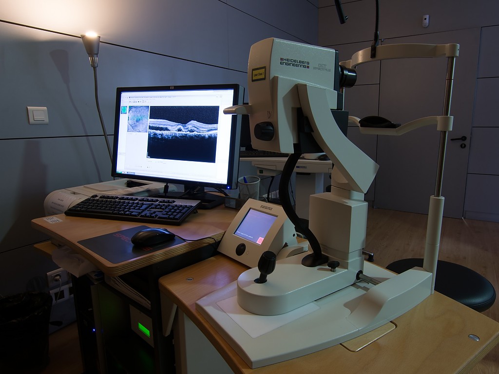
Optical Coherence Tomography - OCT
Optical coherence tomography, or just OCT, is a diagnostic and monitoring technique that allows us to study histological sections of the retina in vivo.
It is a non-invasive, easy-to-perform test, that requires no eye contact and has no side effects. The operation of the OCT is based on the emission of light. The composition of the image is obtained through the difference between the light emitted and the light captured, allowing in this way to make several "cuts" that allow us to visualize the anatomical structures.
In the ocular tomography or OCT, the obtained images are in high resolution. This examination was a great advance in the study of structures of the posterior pole of the eye (retina, vitreous and optic nerve), being very useful in the diagnosis, surgical approach and follow-up of several pathologies:
- macular degenerations;
- pathologies of the vitreoretinal interface (macular hole; epiretinal membrane);
- retinal dystrophies (hereditary diseases);
- glaucoma.
The OCT also provides images of the central retinal structure (fovea) responsible for maximum visual acuity, providing objective information about its microscopic anatomy and its qualitative and quantitative changes.
Preparation:
Usually no relevant preparation required.